Showing posts with label medical. Show all posts

Sunday, March 11, 2018

Dollars for Docs

Pharmaceutical and medical device companies are now required by law to release details of their payments to a variety of doctors and U.S. teaching hospitals for promotional talks, research and consulting, among other categories. Use this tool [link below] to search for general payments (excluding research and ownership interests) made from August 2013 to December 2015.


The following link includes payments from 2013 to 2015

Dollars for Docs

The following link includes payments made through 2016:

Open Payments Data

Other Links:

About the Dollars for Docs Data
“ProPublica’s Dollars for Docs database contains payments to doctors and teaching hospitals from pharmaceutical and medical device companies made between August 2013 and December 2015. The disclosures were required under the Physician Payments Sunshine Act, a part of the 2010 Affordable Care Act.

The database includes “general payments” — 15 categories including promotional speaking, consulting, meals, travel and royalties. It does not include research payments nor does it include physicians’ ownership stakes in companies. Research payments will be included in Dollars for Docs in the future. Detailed descriptions of the payments can be found here.

The doctors included in our tool include medical doctors (MD), dentists, osteopaths (DO), optometrists, podiatrists and chiropractors. The tool does not include nurse practitioners and physician assistants (because companies are not required to report payments to them.) The tool also allows you to search teaching hospitals.

We’ve taken the payment reports, which were released by the Centers for Medicare and Medicaid Services, and compiled them into a single, comprehensive database that allows patients to search for their physician and receive a listing of all payments matching that name. We provide rankings for each doctor to allow comparisons to peers in the same specialty and state.

Our analysis found that most doctors take payments, and that doctors who receive payments are, on average, more likely to prescribe a higher percentage of brand-name drugs. For each physician in Dollars for Docs, we document the number of payments he or she received, the total of those payments, and the number of different companies that paid him or her.

The bulk of each physician’s page is taken up by information on each payment he or she received, the company making the payment, the date of the payment, the names of the drugs and medical devices associated with the payment, and whether the payment was made to a third party entity. Sometimes, payments are not made directly to doctors but instead are provided to their universities, medical practices or research centers. We also note whether a doctor has disputed the payment.

By default, a physician’s page shows aggregate amounts received by year and details of payments in the most recent year reported. Users can display other years by using the dropdown at the top of each doctor page. Company and product pages display aggregate totals across all payment years (2013 to 2015).

Sometimes, more than one company makes payments related to a single drug or device. On each product page, we note the number of companies making payments, as well as the names of those companies. Each payment can also be attributed to more than one product, so we note the percentage of payments that relate only to that product (meaning no other products were mentioned in the payment).
Use the Data

Get the data that powers this investigation. A digital download is available for purchase in the Data Store.

ProPublica has published Dollars for Docs since 2010, at first using payment reports that certain companies were required to publish as part of legal settlements with the federal government. Often, these settlements were related to whistleblower lawsuits alleging improper marketing or kickbacks by the company. The now-archived version of our database includes $4 billion in payments from 17 companies, from 2009 to 2013. An archive of those payments is still available at projects.propublica.org/d4d-archive.

We have made some effort to normalize the data and eliminate duplicates, but data is primarily as it has been reported by the companies to the Centers for Medicare and Medicaid Services.

About Open Payments
“Open Payments is a national transparency program that collects and publishes information about financial relationships between the health care industry (i.e. drug and device companies) and providers (i.e. physicians and teaching hospitals). These relationships may involve payments to providers for things such as research, meals, travel, gifts, or speaking fees. One of the ways that the Centers for Medicare & Medicaid Services (CMS) provides data to the public is through this search tool, which allows the public to search for physicians and teaching hospitals receiving payments, as well as companies that have made payments.

The purpose of the program is to provide the public with a more transparent healthcare system. All information available on the Open Payments database is open to personal interpretation and if there are questions about the data, healthcare consumers should speak directly to the healthcare provider for a better understanding. More information about the program can be found on the CMS Open Payments website.

There you can get an overview of the data that is collected and displayed and learn more about what is included in the data. For other Open Payments related questions, contact the Open Payments team at openpayments@cms.hhs.gov.

Read more...

Friday, April 14, 2017

Natural Cancer Bullets A - Z

Natural cancer bullets A - Z continued:


β-Elemene

Source: Ginger Root


Antitumor effect of β-elemene in non-small-cell lung cancer cells is mediated via induction of cell cycle arrest and apoptotic cell death
“ Beta-elemene is a novel anticancer drug, which was extracted from the ginger plant. However, the mechanism of action of beta-elemene in non-small-cell lung cancer (NSCLC) remains unknown. Here we show that beta-elemene had differential inhibitory effects on cell growth between NSCLC cell lines and lung fibroblast and bronchial epithelial cell lines. ...These data indicate that the effect of beta-elemene on lung cancer cell death may be through a mitochondrial release of the cytochrome c-mediated apoptotic pathway.

Elemene displays anti-cancer ability on laryngeal cancer cells in vitro and in vivo
“ Elemene inhibited the growth of HEp-2 cells in vitro in a dose- and time-dependent manner with an IC50 of 346.5 μM (24 h incubation). Increased apoptosis was observed in elemene-administered cells. Elemene is suspected to enhance caspase-3 activity, and thus inhibit protein expression of eIFs (4E, 4G), bFGF, and VEGF. In vivo, the growth of HEp-2 cell-transplanted tumors in nude mice was inhibited by intraperitoneal injection of elemene. Compared with control groups, elemene significantly inhibited the protein expression of eIFs (4E and 4G), bFGF, and VEGF and decreased the MVD. Conclusions: Elemene inhibits the growth of HEp-2 cells in vitro and in vivo. These data provide useful information for further clinical study on the treatment of LSCC by elemene.

Antiproliferative effect of β-elemene in chemoresistant ovarian carcinoma cells

In this study, we show that beta-elemene inhibited the proliferation of cisplatin-resistant human ovarian cancer cells and their parental cells, but had only a marginal effect in human ovary cells, indicating differential inhibitory effects on cell growth between ovarian cancer cells and normal ovary cells.
Effect of Local Arterial Infusion of β_elemene on Breast Cancer Tissue Inhibition and Cell Apoptosis and Proliferation
“ The effect of local arterial infusion of β_elemene on breast cancer tissue inhibition and cell apoptosis and proliferation was observed.
N-(beta-Elemene-13-yl)tryptophan methyl ester induces apoptosis in human leukemia cells
“ Beta-elemene is an active component of herb medicine Curcuma Wenyujin and N-(beta-elemene-13-yl)tryptophan methyl ester (ETME) was synthesized for increasing its antitumor activity. ETME induced apoptosis in human leukemia HL-60 and NB4 cells at concentrations less than 40 microM. The apoptosis induction ability of ETME was associated with the production of hydrogen peroxide (H(2)O(2)), the decrease of mitochondrial membrane potential, and the activation of caspase-3 that was blocked by catalase. ETME in combination with arsenic trioxide (As(2)O(3)), an agent used to treat acute promyelocytic leukemia, synergistically induced apoptosis in both cell lines by enhanced production of H(2)O(2). These data suggest that ETME induces apoptosis and synergizes with As(2)O(3) in leukemia cells through a H(2)O(2)-dependent pathway.

ß-Glucan

Foods: Mushrooms, Grains & Yeast.
Mushrooms: Shiitake (Lentinula edodes), Reishi (Ganoderma lucidum & Ganoderma tsugae), Maitake (Grifola frondosa), Oyster Mushroom (Pleurotus ostreatus), Cauliflower Mushroom (Sparassis). Grains: Oats, Barley.


Fights cancer and eases the effects of radiation.

NOTE: Literally all of the listed mushrooms above have other important anti-cancer effects not necessarily specified here


ß-Glucan...Uses Antibodies to Target Tumors for Cytotoxic Recognition by Leukocyte Complement Receptor Type 3

“ ß-Glucans were identified 36 years ago as a biologic response modifier that stimulated tumor rejection. In vitro studies have shown that ß-glucans bind to a lectin domain within complement receptor type 3 (CR3; known also as Mac-1, CD11b/CD18, or Mß2-integrin, that functions as an adhesion molecule and a receptor for factor I-cleaved C3b, i.e., iC3b) resulting in the priming of this iC3b receptor for cytotoxicity of iC3b-opsonized target cells. This investigation explored mechanisms of tumor therapy with soluble ß-glucan in mice. Normal mouse sera were shown to contain low levels of Abs reactive with syngeneic or allogeneic tumor lines that activated complement, depositing C3 onto tumors. Implanted tumors became coated with IgM, IgG, and C3, and the absent C3 deposition on tumors in SCID mice was reconstituted with IgM or IgG isolated from normal sera. Therapy of mice with glucan- or mannan-rich soluble polysaccharides exhibiting high affinity for CR3 caused a 57–90% reduction in tumor weight. In young mice with lower levels of tumor-reactive Abs, the effectiveness of ß-glucan was enhanced by administration of a tumor-specific mAb, and in SCID mice, an absent response to ß-glucan was reconstituted with normal IgM or IgG. The requirement for C3 on tumors and CR3 on leukocytes was highlighted by therapy failures in C3- or CR3-deficient mice. Thus, the tumoricidal function of CR3-binding polysaccharides such as ß-glucan in vivo is defined by natural and elicited Abs that direct iC3b deposition onto neoplastic cells, making them targets for circulating leukocytes bearing polysaccharide-primed CR3. Therapy fails when tumors lack iC3b, but can be restored by tumor-specific Abs that deposit iC3b onto the tumors.

Yeast ß-Glucan Amplifies Phagocyte Killing of iC3b-Opsonized Tumor Cells

“ Anti-tumor mAbs hold promise for cancer therapy, but are relatively inefficient. Therefore, there is a need for agents that might amplify the effectiveness of these mAbs. One such agent is -glucan, a polysaccharide produced by fungi, yeast, and grains, but not mammalian cells. -Glucans are bound by C receptor 3 (CR3) and, in concert with target-associated complement fragment iC3b, elicit phagocytosis and killing of yeast. -Glucans may also promote killing of iC3b-opsonized tumor cells engendered by administration of anti-tumor mAbs. In this study, we report that tumor-bearing mice treated with a combination of -glucan and an anti-tumor mAb show almost complete cessation of tumor growth.

Chemosensitization of Carmustine with Maitake β-Glucan on Androgen-Independent Prostatic Cancer Cells
“ This study demonstrates a sensitized cytotoxic effect of BCNU with β-glucan in PC-3 cells, which was associated with a drastic (~80%) inactivation of Gly-I. Therefore, the BCNU/β-glucan combination may help to improve current treatment efficacy by targeting Gly-I, which appears to be critically involved in prostate cancer viability.

Beta glucan induces proliferation and activation of monocytes in peripheral blood of patients with advanced breast cancer

“ Glucans are glucose polymers that constitute a structural part of fungal cell wall. They can stimulate the innate immunity by activation of monocytes/macrophages. In human studies it has been shown that beta glucan has an immunomodulatory effect and can increase the efficacy of the biological therapies in cancer patients. In this prospective clinical trial we assessed in vivo effects of short term oral beta glucan administration on peripheral blood monocytes and their expression of activation markers in patients with advanced breast cancer. ...Oral beta glucan administration seems to stimulate proliferation and activation of peripheral blood monocytes in vivo in patients with advanced breast cancer.

ß-Hydroxyisovalerylshikonin


Source: Lithospermum (herbs).


ß-Hydroxyisovalerylshikonin Inhibits the Cell Growth of Various Cancer Cell Lines and Induces Apoptosis in Leukemia HL–60 Cells
“ ß-Hydroxyisovalerylshikonin (ß-HIVS), which was isolated from the plant, Lithosper-mium radix, inhibited the growth of various lines of cancer cells derived from human solid, tumors at low concentrations between 10-8 and 10-6 M. When HL-60 cells were treated with 10-6 M ß-HIVS for 3 h, characteristic features of apoptosis, such as DNA fragmentation, nuclear fragmentation, and activation of caspase-3–like activity, were observed.

β-Hydroxyisovalerylshikonin and Cisplatin Act Synergistically to Inhibit Growth and to Induce Apoptosis of Human Lung Cancer DMS114 Cells

“ beta-Hydroxyisovalerylshikonin (beta-HIVS) and cisplatin (CDDP) had a synergistic growth-inhibitory effect on cultured human small-cell lung carcinoma DMS114 cells, as well as on human leukemia U937 and epidermoid carcinoma A431 cells, while beta-HIVS and CDDP alone at the same respective concentrations had little effect.

Betulin

Source: The bark of Red Alder trees & White Birch trees, and the mushroom Chaga that grows on White Birch.


Anti-Cancer Effect of Betulin on a Human Lung Cancer Cell Line
“ Betulin is a representative compound of Betula platyphylla, a tree species belonging to the Betulaceae family. In this investigation, we revealed that betulin showed anticancer activity on human lung cancer A549 cells by inducing apoptosis and changes in protein expression profiles were observed.

Betulinic Acid Inhibits Prostate Cancer Growth

“ Betulinic acid is a pentacyclic triterpene natural product initially identified as a melanoma-specific cytotoxic agent that exhibits low toxicity in animal models. Subsequent studies show that betulinic acid induces apoptosis and antiangiogenic responses in tumors derived from multiple tissues;
Betulinic acid induces apoptosis in human neuroblastoma cell lines
Neuroblastoma has long been recognized to show spontaneous regression during fetal development and in the majority of stage 4s infants
Apoptotic activity of betulinic acid derivatives on murine melanoma B16 cell line
“ Exposure of B16 cells to betulinic acid, 23-hydroxybetulinic acid and 3-oxo-23-hydroxybetulinic acid caused a rapid increase in reactive oxidative species production and a concomitant dissipation of mitochondrial membrane potential in a dose- and time-dependent manner, which resulted in cell apoptosis, as demonstrated by fluorescence microscopy, gel electrophoresis and flow-cytometric analysis. Cell cycle analysis further demonstrated that both 3-oxo-23-hydroxybetulinic acid and 23-hydroxybetulinic acid dramatically increased DNA fragmentation at the expense of G1 cells at doses as low as 12.5 and 25 microg/ml, respectively, thereby showing their potent apoptotic properties. Our results showed that hydroxylation at the C3 position of betulinic acid is likely to enhance the apoptotic activity of betulinic acid derivatives (23-hydroxybetulinic acid and 3-oxo-23-hydroxybetulinic acid) on murine melanoma B16 cells.

Betulinic acid: A new cytotoxic compound against malignant head and neck cancer cells

“ In two HNSCC cell lines betulinic acid induced apoptosis, which was characterized by a dose-dependent reduction in cell numbers, emergence of apoptotic cells, and an increase in caspase activity. Western blot analysis of the expression of various Bcl-2 family members in betulinic acid–treated cells showed, surprisingly, a suppression of the expression of the proapoptotic protein Bax but no changes in Mcl-1 or Bcl-2 expression.

Blueberries


Evidence for anti-cancer properties of blueberries: a mini-review.
“ Blueberries are amongst the most commonly consumed berries in the United States. Berries in general are rich in phenolic compounds, which are known for their high antioxidant capacity. Specifically, evidence from in vitro, in vivo and a few clinical studies suggest that blueberries and their active constituents show promise as effective anti-cancer agents, both in the form of functional foods and as nutritional supplements. Some of the mechanisms by which blueberries have been shown to prevent carcinogenesis include inhibition of the production of pro-inflammatory molecules, oxidative stress and products of oxidative stress such as DNA damage, inhibition of cancer cell proliferation and increased apoptosis. This review will focus on the preclinical and clinical evidence that supports blueberries as an anti-cancer fruit, as well as expressing the need for more preclinical studies and the conduction of clinical studies with respect to the cancer preventive ability of blueberries.

Effect of Anthocyanin Fractions from Selected Cultivars of Georgia-Grown Blueberries on Apoptosis and Phase II Enzymes

“ The response correlated positively with dose. The QR activity was lower in all cells treated with an anthocyanin fraction from Tifblue, Powderblue, Brightblue, and Brightwell cultivars than in control cells (P < 0.05). The activity decreased gradually when treated with increased concentrations of anthocyanin fractions (50−150 μg/mL) in the Tifblue and Powderblue cultivars. The GST activity was lower (P < 0.05) in cells treated with anthocyanin fractions from all of the cultivars and at all concentrations. These results indicated that apoptosis was confirmed in HT-29 cells when treated with anthocyanins from blueberry cultivars at 50−150 μg/mL concentrations, but these same concentrations decrease QR and GST activities rather than induce them.
Availability of blueberry phenolics for microbial metabolism in the colon and the potential inflammatory implications
“ Blueberries are a rich source of phenylpropanoid-derived phytochemicals, widely studied for their potential health benefits. Of particular interest for colonic health are the lower molecular weight phenolic acids and their derivatives, as these are the predominant phenolic compounds detected in the colon. Blueberries contained a wide variety of phenolic acids, the majority of which (3371.14 ± 422.30 mg/kg compared to 205.06 ± 45.34 mg/kg for the free phenolic acids) were attached to other plant cell-wall components and therefore, likely to become available in the colon. Cytokine-induced stimulation of the inflammatory pathways in colon cells was four-fold up-regulated in the presence of the free phenolic acid fraction. Incubation of the bound phenolic acids with human faecal slurries resulted in qualitative and quantitative differences in the phenolic compounds recovered. The metabolites obtained by incubation with faecal slurries from one volunteer significantly decreased (1.67 ± 0.69 ng/cm3) prostanoid production, whereas an increase (10.78 ± 5.54 ng/cm3) was obtained with faecal slurries from another volunteer. These results suggest that any potential protective effect of blueberry phenolics as anti-inflammatory agents in the colon is a likely result of microbial metabolism. Studies addressing a wide-range of well-characterised human volunteers will be required before such health claims can be fully established.

Broccoli

Relatives: Romanesco, Broccoflower, Cauliflower, Kale, Raab, Brussel Sprouts, Cabbage, Collards and Kohl Rabi.


Natural Compound in Broccoli Slows Breast Cancer Stem Cells
“ In lab studies, when breast cancer cells were exposed to sulforaphane extract from broccoli, the growth of cancer stems cells slowed down and tumors shrank. The researchers speculate about the possible use of sulforaphane extract to prevent as well as treat breast cancer, someday.
Broccoli Sprouts contain 3X the amount of Sulforaphane Glucosinolate
“Johns Hopkins University researchers found that young broccoli sprouts, in particular, contained high concentrations of SGS. The scientists believe that SGS boosts the body's own antioxidant defense system, including Phase 2 detoxification enzymes, which promote long-lasting antioxidant activity in the body.

Diindolylmethane (DIM)
“At the University of California at Berkeley, the Chairman of the Nutritional Sciences Department and the Director of the National Institutes of Health Cancer Research Program were studying the biological properties of Diindolylmethane (DIM), a naturally occurring compound found in Brassica vegetables (broccoli, cauliflower, cabbage, kale, brussels sprouts), when they made a remarkable discovery: DIM is a potent activator of the immune response system. They patented their discovery and ActivaMune was launched as a first-in-class nutritional supplement to enhance the immune system and support multiple organs throughout the body: breast, prostate, cardiovascular, vision, skin and colon health. ActivaMune's unique and patented formula combines multiple nutrients for maximum effectiveness: Diindolylmethane (DIM), Sulforaphane, Selenium, Lycopene, Lutein, Zeaxanthin, Calcium and Vitamins C, D3 & E.

Surprising Discovery Reveals How Broccoli Fights Cancer


“Broccoli — and to an even greater degree broccoli sprouts — has gained a reputation as a potent cancer-fighter, and recent research sheds new light on the actual mechanics behind its chemoprotective abilities.

One of the compounds in broccoli known to have anti-cancer activity is sulforaphane, a naturally occurring organic sulfur.

Studies have shown sulforaphane supports normal cell function and division while causing apoptosis (programmed cell death) in colon,1 prostate,2 breast3 and tobacco-induced lung cancer4 cells, and reducing the number of cancerous liver tumors in mice.5

Three servings of broccoli per week may reduce your risk of prostate cancer by more than 60 percent.6 Now, Oregon State University (OSU) researchers believe they've discovered yet another way sulforaphane works to protect you against cancer.


Read more...

Natural Cancer Bullets A - Z


Caffeic Acid

Sources: Sweetpotato Leaves, Propolis, Apples, White Grapes, White Wine, Olives, Olive Oil, Spinach, Cabbage, Turnips, Radish, Cauliflower, Bok Choy, Arugula, Kale, Asparagus, and Coffee. 


Growth Suppression of Human Cancer Cells by Polyphenolics from Sweetpotato (Ipomoea batatas L.) Leaves
“Sweetpotato leaves (Ipomoea batatas L.) contain a high content of polyphenolics that consist of caffeic acid, chlorogenic acid, 3,4-di-O-caffeoylquinic acid, 3,5-di-O-caffeoylquinic acid, 4,5-di-O-caffeoylquinic acid, and 3,4,5-tri-O-caffeoylquinic acid. We investigated the suppression of the proliferation of selected human cancer cells by phenolic compounds isolated from sweetpotato leaf. ...Growth suppression of HL-60 cells by 3,4,5-tri-O-caffeoylquinic acid was determined to be the result of apoptotic death of the cells. These results indicate that 3,4,5-tri-O-caffeoylquinic acid may have potential for cancer prevention.
Caffeic acid phenethyl ester induces mitochondria-mediated apoptosis in human myeloid leukemia U937 cells
“Caffeic acid phenyl ester (CAPE), a biologically active ingredient of propolis, has several interesting biological properties including antioxidant, anti-inflammatory, antiviral, immunostimulatory, anti-angiogenic, anti-invasive, anti-metastatic and carcinostatic activities. Recently, several groups have reported that CAPE is cytotoxic to tumor cells but not to normal cells. In this study, we investigated the mechanism of CAPE-induced apoptosis in human myeloid leukemia U937 cells. Treatment of U937 cells with CAPE decreased cell viability in a dose-dependent and time-dependent manner.

Caffeic Acid Phenethyl Ester Induces Apoptosis by Inhibition of NFκB and Activation of Fas in Human Breast Cancer MCF-7 Cells

“Our findings demonstrate that NFκB inhibition is sufficient to induce apoptosis and that Fas activation plays a role in NFκB inhibition-induced apoptosis in MCF-7 cells.

The antioxidant caffeic acid phenethyl ester induces apoptosis associated with selective scavenging of hydrogen peroxide in human leukemic HL-60 cells

“These results suggest that apoptosis induced by CAPE is associated with mitochondrial dysfunction, GSH depletion and selective scavenging of H2O2 in human leukemic HL-60 cells.
Effect of caffeic acid phenethyl ester on proliferation and apoptosis of colorectal cancer cells in vitro
“After HCT116 cells were exposed to CAPE (80, 40, 20, 10, 5, and 2.5 mg/L) for 24, 48, 72, 96 h, CAPE displayed a strong growth inhibitory effect in a dose- and time-dependent manner against HCT116 cells. FCM analysis showed that the ratio of G(0)/G(1) phase cells increased, S phase ratio decreased and apoptosis rate increased after HCT116 cells were exposed to CAPE (10, 5, and 2.5 mg/L) for 24 h. CAPE treatment was associated with decreased cytoplasmic beta-catenin, nuclear beta-catenin and a concurrent increase in beta-catenin protein expression at cell-cell junctions.

Capsaicin

Foods: Hot Peppers.

Sources: Pepper Spray (It's probably a very bad idea to try and breath this stuff). Capsaicin (what makes peppers "hot") has been proven to trigger apoptosis in multiple lines of cancer.


Capsaicin Displays Anti-Proliferative Activity against Human Small Cell Lung Cancer in Cell Culture and Nude Mice Models via the E2F Pathway
“BrdU assays and PCNA ELISAs showed that capsaicin displays robust anti-proliferative activity in four human SCLC cell lines. Furthermore, capsaicin potently suppressed the growth of H69 human SCLC tumors in vivo as ascertained by CAM assays and nude mice models. The second part of our study attempted to provide insight into molecular mechanisms underlying the anti-proliferative activity of capsaicin. We found that the anti-proliferative activity of capsaicin is correlated with a decrease in the expression of E2F-responsive proliferative genes like cyclin E, thymidylate synthase, cdc25A and cdc6, both at mRNA and protein levels.

Capsaicin-induced cell death in a human gastric adenocarcinoma cell line
“Capsaicin, a pungent ingredient found in red pepper, has long been used in spices, food additives, and drugs. Cell death induced by the binding of capsaicin was examined in a human gastric adenocarcinoma cell line (AGS cells).
...Recently, a series of studies have demonstrated that capsaicin inhibits mutagenicity and DNA binding of some chemical carcinogens, possibly by suppressing their metabolic activation[16-18]. With cells in culture, capsaicin-inhibited proliferation of HeLa, ovarian carcinoma, and mammary adenocarcinoma by decreasing NADH oxidase activity[19]. Capsaicin can also alter the expression of tumor forming-related genes by mediating the overexpression of p53 and/or c-myc genes in a Korean stomach cancer cell line[20]. Capsaicin was found to induce apoptosis in T cells by increasing the reactive oxygen species and by a subsequent mitochondrial ransmembrane potential[21]. In this report, we examined the underlying mechanism by which capsaicin induces apoptotic cell death in a human gastric adenocarcinoma cell line (AGS).
Capsaicin-induced apoptosis and reduced release of reactive oxygen species in MBT-2 Murine Bladder Tumor cells
“Capsaicin, the major pungent ingredient in genusCapsicum, has recently been tried as an intravesical drug for overactive bladder and it has also been shown to induce apoptotic cell death in many cancer cells. In this study, we investigated the apoptosis-inducing effect and alterations in the cellular redox state of capsaicin in MBT-2 murine bladder tumor cells. Capsaicin induced apoptotic MBT-2 cell death in a time- and dose-dependent manner. The capsaicin-induced apoptosis was blocked by the pretreatment with Z-VAD-fmk, a broad-range caspase inhibitor, or AcDEVD-CHO, a caspase-3 inhibitor.
Capsaicin-induced apoptosis in human breast cancer MCF-7 cells through caspase-independent pathway
“Our results suggest that capsaicin induces cellular apoptosis through a caspase-independent pathway in MCF-7 cells, and that reactive oxygen species and intracellular calcium ion fluctuation has a minimal role in the process.
Capsaicin Shows Promise In Inhibiting Growth Of Pancreatic Cancer
“In our study, we discovered that capsaicin fed orally to mice with human pancreatic tumors was an extremely effective inhibitor of the cancer process, inducing apoptosis in cancer cells,” said Sanjay K. Srivastava, Ph.D., lead investigator and assistant professor, department of pharmacology, University of Pittsburgh School of Medicine. “Capsaicin triggered the cancerous cells to die off and significantly reduced the size of the tumors.”
TRPV6 mediates capsaicin-induced apoptosis in gastric cancer cells
“In this study, both gastric cancer and normal epithelial cells were treated with capsaicin and examined for apoptosis by Annexin V binding. Our results showed that capsaicin induces apoptosis in both cells, although cancer cells are more susceptible. This susceptibility is dependent on the availability of TRPV6, a calcium-selective channel protein, as overexpression of TRPV6 in normal cells increased capsaicin-induced apoptosis and knockdown of TRPV6 in cancer cells suppressed this action. Our results further demonstrated that capsaicin increases mitochondrial permeability through activation of Bax and p53 in a JNK-dependent manner.

Capsaicin Mediates Cell Death in Bladder Cancer T24 Cells Through Reactive Oxygen Species Production and Mitochondrial Depolarization
“RESULTS: CAP decreased the viability of T24 cells in a dose-dependent manner without marked apoptosis. CAP induced ROS production and mitochondrial membrane depolarization, thereby inducing cell death, not apoptosis, in T24 cells at a concentration of 100 microM or higher. Furthermore, these effects of CAP could be reversed by capsazepine, the antagonist of transient receptor potential vanilloid type 1 channel. In vivo experiment showed that CAP significantly slowed the growth of T24 bladder cancer xenografts as measured by size (661.80 +/- 62.03 vs 567.02 +/- 43.94 mm(3); P

Chillies could help beat cancer as research finds capsaicin destroys diseased cells

“Scientists say breast, colon, bone and pancreatic cancer cells could be caused to self destruct by the spicy ingredient

Cinnamaldehyde

Food: Cinnamon 


Can Cinnamon be the Silver Bullet for Cancer?
“Surprisingly, one of the potential agents for this role, cinnamon, is readily available at your local Café. Two of the main ingredients of cinnamon, cinnamaldehyde and procyanidins, have been previously shown to have health-beneficial activities, such as antioxidant, antimicrobial, anti-inflammatory, and anti-diabetic activity. Moreover, an extract from cinnamon (CE) was recently shown to have antitumor and anti-angiogenesis activity as well.

In a recent article published in Molecular Carcinogenesis, scientists from the US have showed that treatment with CE results in reduced migration of invasive breast and ovarian cancer cells, accompanied by reduced protein expression levels of both VEGF and HIF-1. In addition, a significant suppression of blood-vessel formation (see Figure below) and tumor growth was shown after CE treatment in a human ovarian tumor mouse model, which is the most deadly malignancy in women.
Cinnamon Compound Curtails Cancer
“The second leading cause of cancer deaths in the US, colorectal cancer is a progressive disease generally with poor prognosis. Consequently, scientists are exploring approaches that may prevent the disease. Previously, Georg Wondrak, from the University of Arizona (Arizona, USA) identified the ground bark of Cinnamomum aromaticum (cassia cinnamon) and Cinnamomum verum (Ceylon cinnamon), as rich dietary sources of cinnamaldehyde – an inducer of Nrf2 (nuclear factor-E2-related factor 2), a master regulator of the cellular antioxidant defense that represents a promising molecular target for colorectal cancer chemoprevention.
Cinnamaldehyde induces apoptosis by ROS-mediated mitochondrial permeability transition in human promyelocytic leukemia HL-60 cells
“Cinnamaldehyde is an active compound isolated from the stem bark of Cinnamomum cassia, a traditional oriental medicinal herb, which has been shown to inhibit tumor cell proliferation. In this study, we investigated the effects of cinnamaldehyde on the cytotoxicity, induction of apoptosis and the putative pathways of its actions in human promyelocytic leukemia cells. ...Taken together, our data indicate that cinnamaldehyde induces the ROS-mediated mitochondrial permeability transition and resultant cytochrome c release. This is the first report on the mechanism of the anticancer effect of cinnamaldehyde.
More Cinnamon, Less Cancer
“It has been know for a few years that several types of spices contain large amounts of cancer-fighting compounds that could help in the prevention of the disease. In addition to the well-known health properties of tumeric and ginger, a recent study has suggested that cinnamon could be equally useful in reducing tumour growth by blocking the formation of new vessels using a process called angiogenesis.
Cinnamon extract induces tumor cell death through inhibition of NFκB and AP1
“Cinnamon extract strongly inhibited tumor cell proliferation in vitro and induced active cell death of tumor cells by up-regulating pro-apoptotic molecules while inhibiting NFκB and AP1 activity and their target genes such as Bcl-2, BcL-xL and survivin. Oral administration of cinnamon extract in melanoma transplantation model significantly inhibited tumor growth with the same mechanism of action observed in vitro.

Citral

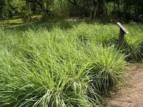
Food: Lemon Grass
Sources: Asian grocery's (lemon grass), Health food stores (extracts).


Fresh lemon grass fields in Israel become Mecca for cancer patients

“A drink with as little as one gram of lemon grass contains enough citral to prompt cancer cells to commit suicide in the test tube. Israeli researchers find way to make cancer cells self-destruct - Ben Gurion University

Lemongrass Fights More than a Dozen Different Types of Cancers

“A 2009 study published in the journal Chemico-Biological Interactions revealed that lemongrass essential oil is effective in targeting at least 12 different human cancer cell lines. Animal trials show that direct injection of lemongrass essential oil inhibits cancer tumors in a dose-dependent way, meaning the higher the dose of the oil, the better the outcome.

When administered at a dosage of 200 milligrams per kilogram (mg/kg) concentration, lemongrass essential oil was shown to inhibit both ascitic (in the fluid of the abdominal cavity) and solid tumors of Ehrlich Ascites (a type of tumor cell line) by 97.34 percent and 57.83 percent, respectively. For Sarcoma-180 tumor cells, the same dosage resulted in ascitic and solid tumor inhibition of 94.07 percent and 36.97 percent, respectively.

An essential oil and its major constituent isointermedeol induce apoptosis by increased expression of mitochondrial cytochrome c and apical death receptors in human leukaemia HL-60 cells.
“An essential oil from a lemon grass variety of Cymbopogon flexuosus (CFO) and its major chemical constituent sesquiterpene isointermedeol (ISO) were investigated for their ability to induce apoptosis in human leukaemia HL-60 cells because dysregulation of apoptosis is the hallmark of cancer cells. ...The easy and abundant availability of the oil combined with its suggested mechanism of cytotoxicity make CFO highly useful in the development of anti-cancer therapeutics.
Citral is a New Inducer of Caspase-3 in Tumor Cell Lines

“Citral, 3,7-dimethyl-2,6-octadien-1-al, a key component of the lemon-scented essential oils extracted from several herbal plants such as lemon grass (Cymbopogon citratus), melissa (Melissa officinalis), verbena (Verbena officinalis) is used as a food additive and as a fragrance in cosmetics. In this study, we investigated the anti-cancer potential of citral and its mode of action. Concentrations of 44.5 μM, comparable to the concentration of citral in a cup of tea prepared from 1 g of lemon grass, induced apoptosis in several hematopoietic cancer cell lines. Apoptosis was accompanied by DNA fragmentation and caspase-3 catalytic activity induction. Citral activity (22.25 μM) was compared to a reference compound like staurosporine (0.7 μM), in respect to DNA fragmentation and caspase-3 enzymatic activity. The apoptotic effect of citral depended on the α,β-unsaturated aldehyde group.

Citral inhibits cell proliferation and induces apoptosis and cell cycle arrest in MCF-7 cells.
“In this study, we investigated the effect of citral (3,7-dimethyl-2,6-octadienal), a key component of essential oils extracted from several herbal plants, on the proliferation rate, cell cycle distribution, and apoptosis of the human breast cancer cell line MCF-7. The effects of this compound were also tested on cyclo-oxygenase activity. Citral treatment caused inhibition of MCF-7 cell growth (IC(50)-48 h: 18 x 10(-5)m), with a cycle arrest in G(2)/M phase and apoptosis induction. Moreover, we observed a decrease in prostaglandin E(2) synthesis 48 h after citral treatment. These findings suggest that citral has a potential chemopreventive effect.

Cod Liver Oil

Women and Lung Cancer - Could Cod Liver Oil Improve Survival
“My Scandinavian, cod-liver-oil-serving grandma was right again – based on the results of a new Norwegian study. Women who used cod liver oil daily for a year prior to their diagnosis of lung cancer, had a 44% lower chance of dying. Though my grandmother would have turned 100 last week, support for her advice will be published next month in the International Journal of Cancer. Researchers looked at questionnaires completed by over 68,000 women between 1996 and 1999. They then analyzed 2,242 women who developed cancer following the questionnaire and up to the year 2007. Women who used cod liver oil daily, had a 23% lower chance of dying from solid tumors in general (breast, colorectal, and lung cancers), and a 44% reduced risk of dying from lung cancer.

Coenzyme Q10


Foods: Fish, chicken, peanuts, seasame seeds, pistachios, olive oil, soy beans, grapeseed, parsley, perilla, broccoli (and relatives).


How does CoQ10 work against cancer?
“Co-Q10 is present in most "eukaryotic" cells (muscle tissues and more). It's produced naturally in the body, which uses it for cell growth and to protect cells from damage that could lead to cancer. In animals it's most concentrated in the heart. Co-Q10 both boosts the immune system, and has been shown to induce apoptosis.
A Gentle Cancer Killer
“In laboratory and animal studies, the UM researchers found that by delivering CoQ10 to cancer cells and tissues, the molecule induced apoptosis, which is the normal programmed cell death that goes awry in the disease process.
Coenzyme Q10 (PDQ®)–Patient Version
Low blood levels of coenzyme Q10 have been found in patients with myeloma, lymphoma, and cancers of the breast, lung, prostate, pancreas, colon, kidney, and head and neck. Studies suggest that coenzyme Q10 may help the immune system work better. Partly because of this, coenzyme Q10 is used as adjuvant therapy for cancer. Adjuvant therapy is treatment given following the primary treatment to increase the chances of a cure.
“Clinical trials have shown that coenzyme Q10 helps protect the heart from the damaging side effects of doxorubicin, a drug used to treat cancer.

Normalizattion of BCL-2 family members in breast cancer by Coenzyme Q10
“Because of their integral role in intrinsic apoptosis any imbalance can lead to a variety of diseases; under expression can lead to degenerative diseases while over expression can lead to cancer and autoimmune disease. Due to their life or death role in the cell, Bcl-2 family members are currently the targets of many therapies in various disease states. Bcl-2 itself is over expressed in most tumors and all anti-apoptotic Bcl-2 family members are considered to have oncogenic potential. Conversely, the pro-apoptotic members are considered to be tumor suppressors and many mimetics are foci for cancer research. ...Both pro- and anti-apoptotic protein levels were measured in the two breast cancer cell lines after Q10 exposure. Protein levels were measured at 4,8,12, and 24 hours respectively in order to capture evidence of Q10's normalizing influence on disrupted apoptotic function. In the MCF-7 cell line Bcl-2 levels were seen to significantly drop after only 4 hours of Q10 exposure.

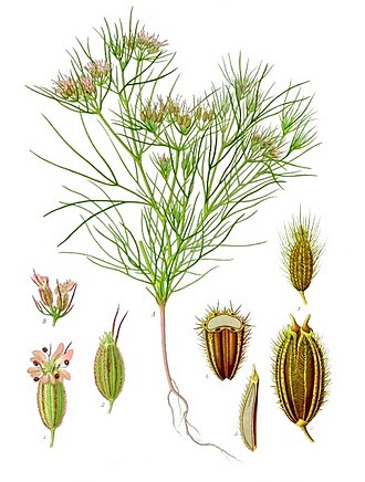
Cuminum

Food: Cumin Seeds.

Primary ingredient in Chili Powder.

Cumin: natures potent cancer combatant
“This herb has been seen to effectively decrease the incidence of chemically induced tumors of the stomach, colon, and cervix. Its significant antioxidant activity and the ability to modulate the metabolism of carcinogens (toxins) explain its cancer-preventive prowess. Cumin seeds are known to induce the activity of glutathione-S-transferase, a protective enzyme that helps eliminate cancer-causing substances. Cumin offers a significant level of caffeic, chlorogenic, ferulic, and other phenolic acids that have anti-inflammatory potential.

Curcumin

Foods: Tumeric roots / powder, curry powder, yellow mustard.
Sources: Health food stores (extracts), large farmers markets (roots), Indian grocery stores (tumeric powder in bulk).


Induction of apoptosis in human lung cancer cells by curcumin
“This study investigated the cellular and molecular changes induced by curcumin leading to the induction of apoptosis in human lung cancer cell lines—A549 and H1299. A549 is p53 proficient and H1299 is p53 null mutant. The lung cancer cells were treated with curcumin (0–160 μM) for 12–72 h. Curcumin inhibited the growth of both the cell lines in a concentration dependent manner. Growth inhibition of H1299 cell lines was both time and concentration dependent. Curcumin induced apoptosis in both the lung cancer cell lines. A decrease in expression of p53, bcl-2, and bcl-XL was observed after 12 h exposure of 40 μM curcumin. Bak and Caspase genes remained unchanged up to 60 μM curcumin but showed decrease in expression levels at 80–160 μM. The data also suggest a p53 independent induction of apoptosis in lung cancer cells.

Curcumin induces apoptosis in immortalized NIH 3T3 and malignant cancer cell lines
“Curcumin, which is a widely used dietary pigment and spice, has been demonstrated to be an effective inhibitor of tumor promotion in mouse skin carcinogenesis. We report that curcumin induces cell shrinkage, chromatin condensation, and DNA fragmentation, characteristics of apoptosis, in immortalized mouse embryo fibroblast NIH 3T3 erb B2 oncogene-transformed NIH 3T3, mouse sarcoma S180, human colon cancer cell HT-29, human kidney cancer cell 293, and human hepatocellular carcinoma Hep G2 cells

Curcumin induces apoptosis in human breast cancer cells
“The aim of this study was to determine the mechanisms of curcumin-induced human breast cancer cell apoptosis. From quantitative image analysis data showing an increase in the percentage of cells with a sub-G0/G1 DNA content, we demonstrated curcumin-induced apoptosis in the breast cancer cell line MCF-7, in which expression of wild-type p53 could be induced. Apoptosis was accompanied by an increase in p53 level as well as its DNA-binding activity followed by Bax expression at the protein level. Further experiments using p53-null MDAH041 cell as well as low and high p53-expressing TR9-7 cell, in which p53 expression is under tight control of tetracycline, established that curcumin induced apoptosis in tumor cells via a p53-dependent pathway in which Bax is the downstream effector of p53. This property of curcumin suggests that this molecule could have a possible therapeutic potential in breast cancer patients.
Chemopreventive Effect of Curcumin, a Naturally Occurring Anti-Inflammatory Agent, during the Promotion/Progression Stages of Colon Cancer
“The inhibition of adenocarcinomas of the colon was, in fact, dose dependent. Administration of curcumin to the rats during the initiation and postinitiation stages and throughout the promotion/progression stage increased apoptosis in the colon tumors as compared to colon tumors in the groups receiving AOM and the control diet. Thus, chemopreventive activity of curcumin is observed when it is administered prior to, during, and after carcinogen treatment as well as when it is given only during the promotion/progression phase (starting late in premalignant stage) of colon carcinogenesis.

Antiproliferation and apoptosis induced by curcumin in human ovarian cancer cells

“Curcumin, an active ingredient from the rhizome of the plant, Curcuma longa, has antioxidant, anti-inflammatory and anti-cancer activities. It has recently been demonstrated that the chemopreventive activities of curcumin might be due to its ability to inhibit cell growth and induce apoptosis. In the present study, we have investigated the effects of curcumin on growth and apoptosis in the human ovarian cancer cell line Ho-8910 by MTT assay, fluorescence microscopy, flow cytometry and Western blotting. Our data revealed that curcumin could significantly inhibit the growth and induce apoptosis in Ho-8910 cells. A decrease in expression of Bcl-2, Bcl-X(L) and pro-caspase-3 was observed after exposure to 40 microM curcumin, while the levels of p53 and Bax were increased in the curcumin-treated cells. These activities may contribute to the anticarcinogenic action of curcumin.
Curcumin, an antioxidant and anti-tumor promoter, induces apoptosis in human leukemia cells

“Curcumin, widely used as a spice and coloring agent in food, possesses potent antioxidant, anti-inflammatory and anti-tumor promoting activities. In the present study, curcumin was found to induce apoptotic cell death in promyelocytic leukemia HL-60 cells at concentrations as low as 3.5 micrograms/ml.
Notch-1 down-regulation by curcumin is associated with the inhibition of cell growth and the induction of apoptosis in pancreatic cancer cells
“Curcumin inhibited cell growth and induced apoptosis in pancreatic cancer cells. Notch-1, Hes-1, and Bcl-XL expression levels concomitantly were down-regulated by curcumin treatment. These results correlated with the inactivation of NF-κB activity and increased apoptosis induced by curcumin. The down-regulation of Notch-1 by small-interfering RNA prior to curcumin treatment resulted in enhanced cell growth inhibition and apoptosis.

Read more...

Wednesday, April 12, 2017

Second Opinion from Doctor Results in Different Diagnosis 88% of Time


Science Daily clearly takes point of view of the Medical Industrial Complex (MIC)--profit before patients--that the value of second opinions are demonstrated in a study, whereas I see this from the highly skeptical-of-the-deeply-rooted-MIC point of view that it can't be trusted 88% of the time. We don't know whether the first or second opinion is right or wrong. What about a third opinion? Would that opinion disagree with the first and the second?
“Many patients come to Mayo Clinic for a second opinion or diagnosis confirmation before treatment for a complex condition. In a new study, Mayo Clinic reports that as many as 88 percent of those patients go home with a new or refined diagnosis — changing their care plan and potentially their lives. Conversely, only 12 percent receive confirmation that the original diagnosis was complete and correct.
We take it for granted that the Mayo Clinic is right because, well, they're the Mayo Clinic, the poster child for the MIC, this vast networked system that we've been indoctrinated to believe in since birth.
It's not that the MIC is all bad, or hasn't saved many lives and improved the quality of many others.  It's the fact that the opposite is also true, it has destroyed and ruined lives, possibly in equal measure   and we're made to believe there are no other viable options. 
“We are not saying that there are no useful or helpful things within the MIC. It has saved many of our lives or the lives of people we love. We are not anti healthcare or science, but are rather exposing the reality that many of us are dependent on the MIC while we are simultaneously trying to change it and ultimately build alternatives to it. Many of us don’t want to have to turn to the MIC, yet have few other viable options. And still many of us are fighting for access to current (or better) services within the MIC. There are no easy answers and the contradictions we are living in are often painful and unjust. Similar to our work to resist and challenge capitalism or to create alternatives to the police and prisons, resisting and challenging the MIC is rife with complexity and there is so much we need that we don’t have yet.

We are asking, why we have so few options when it comes to our healthcare needs? And why insurance and pharmaceutical companies get to call the shots on the kind of care we receive—or don’t? Why don’t we talk more about the ways that forced medicalization has become part of our prison system? Or how non-western and alternative healing practices are often no less ableist than western medical practices? We are asking, what could “health,” “wellness,” “care,” “accessibility” and “sustainability” look like in practice, outside of theory? We are revealing where and how the MIC is already in our lives in ways we might not have thought of before. We are urging us all to connect the MIC to our political work, because healing, wellness, care, “health” and disability are part of whatever liberatory work we are engaged in.

Links:

Medical Industrial Complex Visual
“The Medical Industrial Complex is an enormous system with tentacles that reach beyond simply doctors, nurses, clinics, and hospitals. It is a system about profit, first and foremost, rather than “health,” well being and care. Its roots run deep and its history and present are connected to everything including eugenics, capitalism, colonization, slavery, immigration, war, prisons, and reproductive oppression. It is not just a major piece of the history of ableism, but all systems of oppression.

Read more...

Monday, July 20, 2015

Solving the Brutal Murder of a Prominent Orthopedic Surgeon and World Renowned Expert in Cancer Research

Dr. Mary Stults Sherman and the apartment building she was found murdered
Prominent orthopedic surgeon and world renowned expert in cancer research,  Dr. Mary Sherman (left) was brutally murdered on July 21, 1964 (the same day the Warren Commission started taking testimony on Lee Harvey Oswald’s activities in New Orleans) in her apartment building on St. Charles Avenue, in New Orleans. This 'assassination' remains officially unsolved--maybe because of the memo from J Edgar Hoover telling SAC-NO (Special Agent-in-charge of the New Orleans FBI office) that no active FBI investigation was to be conducted into the Mary Sherman murder--but there are some investigators and authors, in particular, Edward T. Haslam, author of Dr. Mary’s Monkey: How the Unsolved Murder of a Doctor, a Secret Laboratory in New Orleans and Cancer-Causing Monkey Viruses are Linked to Lee Harvey Oswald, the JFK Assassination and Emerging Global Epidemics, and Mary, Ferrie and the Monkey Virus: Story of an Underground Medical Laboratory, who discovered the truth regarding this very strange murder and, believe it or not, the assassination of John F. Kennedy is closely intertwined. 

Dr. Sherman was stabbed in the heart, liver, arm, leg, and stomach, and her right arm and rib cage were completely incinerated, totally disintegrated, yet the hair on her head remained untouched. The incinerated body parts supposedly occurred due to a small mattress fire, but the investigators determined that the massive burns inflicted upon her could not have occurred in her apartment.  Of course, the details of her missing arm and ribs were not released to the public.  Moreover, the wound to the heart hemorrhaged but the wound to the liver did not, indicating that she was alive at the time of the heart wound but already dead by the time the wound to the liver was inflicted.

Who? What? Why?


A little more history on the subject:

Read more...

Monday, November 18, 2013

Mycoplasma, AIDS, Degenerative Diseases: What You Don't Know Can Kill You.

John D. Rockefeller  Sr.'s merger with pharmaceutical and chemical giant, I.G. Farben (Auschwitz was a 100% subsidiary of IG Farben) in 1928 "created the largest and most powerful cartel the world has ever known. Not only has that cartel survived through the years, it has grown and prospered. Today it plays a major role in both the science and politics of cancer therapy."

It's important to know Big Pharma's "crimes against humanity" history, because that history fostered the chemical-based drug treatment basis for our "orthodox" healthcare system today. The industry claims that their aim is finding medical truth, and that may be true; nevertheless, publicizing that truth, once found, if that truth entails inexpensive solutions or remedies, will not happen for obvious reasons. There is nothing Big Pharma likes more than chronic illness, for which they can produce chronic palliative costly treatments that make the patient chronically dependent on them.

Which brings me to the seldom, if ever, publicized human pathogen: mycoplasmas. They are the smallest free-living bacteria that can assume multiple shapes including round, pear shaped and even filamentous forms because they lack a cell wall. This makes it possible for them to pass through  filters used to remove bacteria; and hide inside the cell from our immune system, as well as from common antibiotics, all the while interfering with the cell machinery. Because mycoplasmas have lost most of their genetic material; a strict dependence on the host for nutrients and refuge determines its ability to survive and grow.

Mycoplasma is the co-factor that alters the human immune system and opens the door for the autoimmune degenerative diseases such as AIDS, malignant transformation, chromosomal aberrations, Chronic Fatigue Syndrome, Gulf War Syndrome (military vaccine), Amyotrophic lateral sclerosis (ALS), often referred to as Lou Gehrig\'s Disease (every single patient shows mycoplasmal infection), Alzheimer’s, Crohns Disease, Parkinson’s, Huntington’s, Lupus, Lyme disease, Fibromyalgia, Rheumatoid Arthritis, Multiple Sclerosis, Type One Diabetes, Autistic Spectrum Disorder, and cancer.  Yet, these pathogenic organisms show up in vaccines.
“Because certain species of the mycoplasma have an absolute growth requirement for the up-take of pre-formed sterols, including cholesterol they can cause the ‘spontaneous degeneration’ of the cells that they invade. If they do not cause sufficient damage to kill the cell, they at least compromise its capacity to defend itself from other disease agents, such as those which present as Kaposi’s sarcoma, pneumoniae carinii pneumonia, lymphadenopathy, and so on.” Donald W. Scott and William L.C. Scott,
“From its inception, the biowarfare program was characterized by continuing in-depth review and participation by the most eminent scientists, medical consultants, industrial experts and government officials, and it was classified Top Secret. The US Public Health Service also closely followed the progress of biological warfare research and development from the very start of the program, and the Centers for Disease Control (CDC) and the National Institutes of Health (NIH) in the United States were working with the military in weaponising these diseases. These are diseases that have existed for thousands of years, but they have been weaponized—which means they’ve been made more contagious and more effective. And they are spreading.” Donald W. Scott
Currently, Dr. Alan Cantwell is one of the most well-known popular proponents of the link between cancer and bacteria. He has written numerous articles and books on the subject after he isolated and reported cell wall deficient bacteria in breast cancer, Kaposi’s sarcoma and Hodgkin’s disease. He states,
”If a disease like cancer is indeed caused by microscopic bacteria, it would indicate physicians have been unable to see what was quite plain for some nineteenth and twentieth century scientists to observe using simple light microscopy."
Eventually, constantly under intense criticism, Cantwell was ostracized by many in the medical profession.

Females are four times more likely to be infected with Mycoplasmas than males. The same ratio of males to females applies to Rheumatoid Arthritis, Fibromyalgia, Chronic Fatigue and other related auto-immune disorders. Men, pound for pound, have 25% more blood; hence, hemoglobin - the protein molecule in red blood cells that carries oxygen from the lungs to the body's tissues - than females.

AIDS and Mycoplasma: The Crime Beyond Belief

Donald W. Scott, editor of The Journal of Degenerative Diseases (pictured above) and co-founder of the Common Cause Medical Research Foundation, reveals the true and hidden story of weaponized mycoplasma and the protracted creation of AIDS by governments and private corporations. part 1 of a series drawn from the 90 min. talk given at the 9th annual conference, Sudbury, Ontario (Aug 29-31, 2008).  JODD (Journal of Degenerative Disease); Box 133 Station B; Sudbury Ontario; Canada P3E4N5  $25/year


Links:

Fluid Mosaic Model Membrane


The Linking Pathogen in Neurosystemic Diseases

Mycoplasmas Stealth Pathogens

Campaign for Truth in Medicine

Mycoplasma Protocol

Institute for Molecular Medicine

Read more...

Wednesday, September 26, 2012

Harvesting Organs Before Death.

I don't think it gets any creepier than this: ripping out body parts before the patient dies.

That's right. The New York Organ Donor Network is accused of pressuring doctors to declare patients dead before they die in order to harvest their organs.

"New York hospitals are routinely "harvesting" body parts from patients before they're even dead, a lawsuit is claiming.
The suit claims that transplant non-profit, The New York Organ Donor Network, of bullying hospital staffers to declare patients brain dead when they are still alive so to take their organs. New York hospitals are routinely "harvesting" body parts from patients before they're even dead, a lawsuit is claiming. The suit claims that transplant non-profit, The New York Organ Donor Network, of bullying hospital staffers to declare patients brain dead when they are still alive so to take their organs. The suit accuses the transplant non-profit, The New York Organ Donor Network, of bullying hospital staffers to declare patients brain dead when they are still alive in order to take their organs.
Plaintiff Patrick McMahon, 50, an Air Force combat veteran, is a former transplant coordinator who claims he was fired just four months into the job for protesting about the practice and estimates that one in five patients is still showing signs of brain activity when surgeons declare them dead and start ripping out their body parts.
"They're playing God," McMahon told New York Post. The lawsuits, filed in Manhattan Supreme Court Tuesday, cited four examples of improper organ harvesting.
One of the examples cited details of a 19-year-old man injured in a car crash who was still struggling to breath and showed signs of brain activity when doctors at Nassau University Medical Center declared him brain dead under pressure from the donor-network officials, including Director Michael Goldstein, who allegedly said during a conference call: "This kid is dead, you got that?" the suit claims.
McMahon said that the teenager could have easily recovered.
"I have been in Desert Storm, Iraq and Afghanistan in combat," he told New York Post. "I worked on massive brain injuries, trauma, gunshot wounds, IEDs. I have seen worse cases than this and the victims recover."
The three other examples of patients who were still clinging to life when doctors declared them brain dead included a female patient admitted to St. Barnabas Hospital, a man admitted to Kings County Hospital in Brooklyn and a woman admitted to Staten Island University Hospital after a drug overdose who was given "a paralyzing anesthetic" because her body was still jerking, according to the Post.
McMahon said when he flagged up the injection, another network employee told hospital personnel McMahon was "an untrained troublemaker with a history of raising frivolous issues and questions," the suit charged. "I had a reputation for raising a red flag," he said.
McMahon accuses the federally-funded network of having a "quota" system and hiring "coaches" to teach staff how to be more persuasive in getting family members to give consent to organ donation. McMahon claims that on November 4, he told the network's CEO and president, Helen Irving, that "one in five patients declared brain dead show signs of brain activity at the time the Note is issued."
However, according to the suit, Irving replied: "This is how things are done.
There’s a lot of money at stake here.  To be sure, when quota systems are in place, and they are, the pressure is on medical professionals to either look the other way, or declare death prematurely.  

Links:

Parents' Hospital Lawsuit Says Teen Was 'Killed' For Organs

Read more...

Friday, March 30, 2012

Woman Arrested After Begging For Care At Hospital, Then Dies In Jail

It is said that all it takes for the triumph of evil is for "good" men to do nothing. Good? How is a man good if he does nothing to help another human being who is suffering? Dying? Especially if that man has taken an oath to watch over the life and health of  their fellow human beings (doctors), or has sworn to protect their fellow human beings (police). 

Sadly, for Anna Brown, a 29-year-old African American woman left homeless after a tornado in 2010 that destroyed her home, after she lost her job at a sandwich shop, met with the most blatant and callous disregard in her time of need, instead. 
Ms. Brown, 29, had sprained her ankle that week and on September 20, went to Saint Louis University Hospital for treatment. The X-rays were negative and she was given painkillers and discharged. But Ms. Brown was not satisfied, sensing something more was wrong with her. She refused to leave the hospital, so police were called. Hospital security called police, so Ms. Brown, in a wheelchair due to the pain in her legs, rowed herself next door to a children’s hospital. Though doctors there said she did in fact have tenderness in her legs, they said they could not treat her because it was a pediatric hospital. Refusing to return to SLU Hospital, an ambulance took her to SSM St. Mary’s Health Center, a hospital with a mission statement emphasizes “special concern for people who are materially poor and vulnerable.” Ms. Brown was homeless and on Medicaid after a tornado in 2010 destroyed her home and she lost her job at a sandwich shop.

Ms. Brown was given ultrasounds on both legs, which did not find any blood clots or other abnormalities, according to State inspectors who examined her medical records. The hospital gave Ms. Brown the number to several homeless shelters and sent her on her way. But Ms. Brown returned eight hours later, now complaining of leg and abdominal pain. Hospital staff refused to treat her, giving her discharge papers which she refused to sign. Richmond Heights Police, who were already at the scene, wheeled Ms. Brown out of the hospital, as she yelled “my legs don’t work!” At the request of the hospital, police arrested Ms. Brown and charged her with trespassing.

When they arrived at the jail, Ms. Brown told the police she could not put pressure on her legs, so they dragged her out of the car by her arms. Police listed “possible drug use” as their reason for Ms. Brown’s behavior, since hospital staff told them she was “fit for confinement.” Though the woman moaned in pain and begged for help, police dragged her into a jail cell and laid her on the concrete floor, even though a cot was right next to her. Ms. Brown stopped moving and breathing fifteen minutes later. Paramedics tried to revive her without success, so they rushed her back to St. Mary’s, where she died within an hour. The autopsy revealed she died from blood clots in her legs that ultimately lodged in her lungs.

Read more...

Sunday, January 15, 2012

Smoking is Good For You?

According to Niels Ipsen, an environmental biologist and Klaus Kjellerup, a researcher who analyzed and summarized 40 years of scientific research. The claim is that nicotine boosts brain performance. They do not address the lungs. I guess breathing isn't as important if your brain's on fire.

After 40 years of scientific research on the effects of nicotine, researchers now say that they have sound scientific proof that smoking and nicotine have a significant positive effect on human brain performance.

The brain works better when it gets nicotine - almost like an optimized computer. Nicotine is a "work-drug" that enables its consumers to focus better and think faster. The brain also becomes more enduring, especially in smokers: Nicotine experiments show that smokers in prolonged working situations are able to maintain concentration for many hours longer than non-smokers.

This seems like a paradox considering the smoking bans imposed on workplaces in many countries - but it is nonetheless the picture emerging from hundreds of scientific studies of smoking and nicotine. It seems very unlikely that companies would be able to stop smoking in workplaces with many smokers without experiencing a decline in labor productivity.

Generally nicotine boosts the brain to work 10-30% more efficiently in a number of areas. This is especially true for smoking - but also true when using smokeless nicotine. But at the same time, when smokers and nicotine users abstain, they experience a perhaps equally great decline in the effect. This is called the "withdrawal effect" - a nicotine craving, especially for smokers.

Thus the difference between smoking and smoking abstinence is very pronounced for a smoker - a difference of perhaps as much as 50%. And, according to the scientists, this answers the question: Why do people smoke? The answer is simple: Because smoking boosts their brain power.
There are over 4000 chemicals (many of them, combustion by-products) in store bought cigarettes that include formaldehyde, arsenic and cyanide.  Now, certain chemicals reduce the tar intake. That's a good thing, right? But, to be sure, there are far more bad things.  For one, cigarette manufacturers like to enhance nicotine delivery to increase likelihood of addiction. For example, adding ammonia enables the smoker to freebase nicotine. Moreover, they add other chemicals to keep the cigarette from extinguishing itself. That can't be good.

And if that's not bad enough, fire safe cigarette legislation,  in all 50 states has produced the Fire Standards Compliant cigarette, which is held together with Ethylene Vinyl Acetate - carpet glue!   If this isn't incentive to roll your own...
Ethylene Vinyl Acetate, the product that holds the cigarette paper together, is essentially carpet glue. The product has no accepted internal (ingestion) purpose, and is considered to be highly toxic. In fact, the Material Safety Data Sheet, the “expert” on material uses says the following about ethylene vinyl acetate, “Not considered hazardous under normal conditions of use.” Normal conditions, of course, are those that include gluing a new carpet to the floor. Yuck!

The Data Sheet continues, in the same paragraph stating; “Can release irritating or toxic fumes and vapors if involved in a fire.” Naturally, all cigarettes are involved in a fire, as that is the only way they can be consumed.

What’s in a FSC?
Well, a lot. The health risks are plenty when inhaling the smoke from Fire Safe Cigarettes. FSC’s produced 13.9% more Naphthalene and 11.4% more carbon monoxide than regular cigarettes. Naphthalene is a nasty chemical, the main ingredient in moth balls, and contributes to the following conditions: headache, nausea, vomiting, diarrhea, malaise, confusion, anemia, jaundice, convulsions, and coma.
...that is, unless you want to try the so-called safe alternative, Hookah or water-pipe smoking, that seems to be on the rise, especially amongst teens. Apparently, some think the smoke is filtered by the water;  hence, is safer than regular cigarette smoking. Not true, "a hookah, which is smoked for about 45 minutes, delivers 36 times more tar than a cigarette, 15 times more carbon monoxide and 70% more nicotine.".  The Mayo Clinic agrees.

In conclusion, while scientists have found that nicotine might be good for you in certain circumstances, and  that smoking might help prevent the onset of various dementias (nicotine), and that smokers, in general, are more likely to avoid obesity, smoking still drastically increases your chances of cancer and heart disease.  

Science is conclusive: Tobacco increases work capacity

Read more...
Iraq Deaths Estimator
Petitions by Change.org|Start a Petition »

  © Blogger templates The Professional Template by Ourblogtemplates.com 2008

Back to TOP